")
Volume 31, Number 4 (2015)
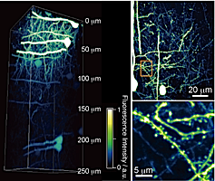
Cover illustration of this month"Multi-point Scanning Two-photon Excitation Microscopy by Utilizing a High-peak-power 1042-nm Laser" by Kohei OTOMO et al. (p.307). See larger image
Hot Articles − Volume 31, Number 4 (2015)
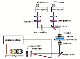
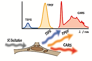
 Multimodal Imaging of Living Cells with Multiplex Coherent Anti-stokes Raman Scattering (CARS), Third-order Sum Frequency Generation (TSFG) and Two-photon Excitation Fluorescence (TPEF) Using a Nanosecond White-light Laser Source
Analytical Sciences, 2015, 31(4), 299.
DOI: 10.2116/analsci.31.299
| ||
|
Multi-point Scanning Two-photon Excitation Microscopy by Utilizing a High-peak-power 1042-nm Laser
Analytical Sciences, 2015, 31(4), 307.
DOI: 10.2116/analsci.31.307
| ||
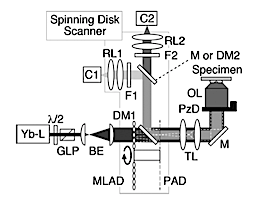
Table of Contents − Volume 31, Number 4 (2015)
Special Issue: Molecular Imaging for Bioanalysis
Guest Editorial
|
“Molecular Imaging for Bioanalysis”
Analytical Sciences, 2015, 31(4), 243.
DOI: 10.2116/analsci.31.243
| ||
Reviews
|
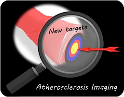
New Targets of Molecular Imaging in Atherosclerosis: Prehension of Current Status
Analytical Sciences, 2015, 31(4), 245.
DOI: 10.2116/analsci.31.245
| ||
|
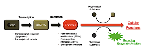
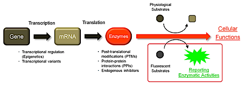
Evaluation of Enzymatic Activities in Living Systems with Small-molecular Fluorescent Substrate Probes
Analytical Sciences, 2015, 31(4), 257.
DOI: 10.2116/analsci.31.257
| ||
|
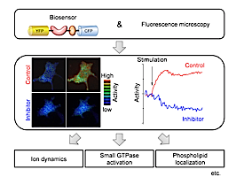
Fluorescent Protein-based Biosensors to Visualize Signal Transduction beneath the Plasma Membrane
Analytical Sciences, 2015, 31(4), 267.
DOI: 10.2116/analsci.31.267
| ||
|
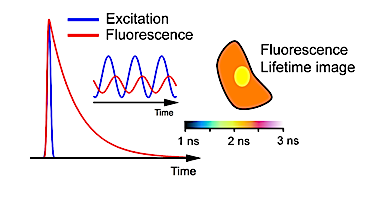
Sensing of Intracellular Environments by Fluorescence Lifetime Imaging of Exogenous Fluorophores
Analytical Sciences, 2015, 31(4), 275.
DOI: 10.2116/analsci.31.275
| ||
|
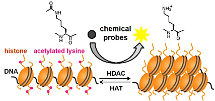
Chemical Tools for Probing Histone Deacetylase (HDAC) Activity
Analytical Sciences, 2015, 31(4), 287.
DOI: 10.2116/analsci.31.287
| ||
|
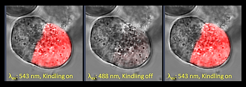
An Efficient Method is Required to Transfect Non-dividing Cells with Genetically Encoded Optical Probes for Molecular Imaging
Analytical Sciences, 2015, 31(4), 293.
DOI: 10.2116/analsci.31.293
| ||
Original Papers
|
Multimodal Imaging of Living Cells with Multiplex Coherent Anti-stokes Raman Scattering (CARS), Third-order Sum Frequency Generation (TSFG) and Two-photon Excitation Fluorescence (TPEF) Using a Nanosecond White-light Laser Source
Analytical Sciences, 2015, 31(4), 299.
DOI: 10.2116/analsci.31.299
| ||
|
Multi-point Scanning Two-photon Excitation Microscopy by Utilizing a High-peak-power 1042-nm Laser
Analytical Sciences, 2015, 31(4), 307.
DOI: 10.2116/analsci.31.307
| ||
|
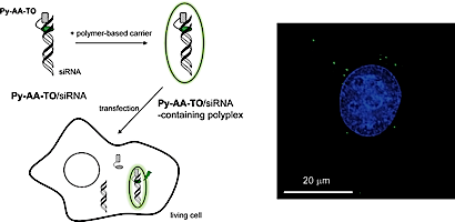
Fluorescence Imaging of siRNA Delivery by Peptide Nucleic Acid-based Probe
Analytical Sciences, 2015, 31(4), 315.
DOI: 10.2116/analsci.31.315
| ||
|
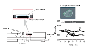
An Enzyme-entrapped Agarose Gel for Visualization of Ischemia-induced L-Glutamate Fluxes in Hippocampal Slices in a Flow System
Analytical Sciences, 2015, 31(4), 321.
DOI: 10.2116/analsci.31.321
| ||
Notes
|
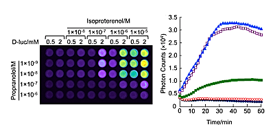
High-throughput Live Cell Imaging and Analysis for Temporal Reaction of G Protein-coupled Receptor Based on Split Luciferase Fragment Complementation
Analytical Sciences, 2015, 31(4), 327.
DOI: 10.2116/analsci.31.327
| ||
|
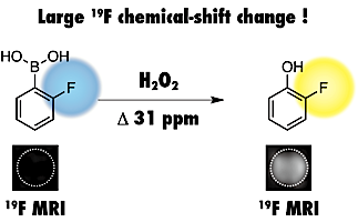
Phenylboronic Acid-based 19F MRI Probe for the Detection and Imaging of Hydrogen Peroxide Utilizing Its Large Chemical-Shift Change
Analytical Sciences, 2015, 31(4), 331.
DOI: 10.2116/analsci.31.331
| ||
Announcements
|
Analytical Sciences, 2015, 31(4), 337.
DOI: 10.2116/analsci.31.337
| ||